 |
| Cortical thickness color maps for the outer table of the two cadaveric skulls A and B using the cortical density method with microCT high resolution thickness of samples overlaid. Source |
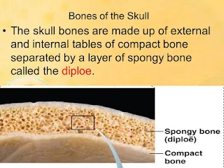
The skull is a composite of a spongy material sandwiched by two layers of hard, strong, brittle bone. The spongy middle is called diploe while the hard layers are called the inner and outer tables. This structure provides a compromise of minimum mass, the ability to carry point loads and impact resistance.
The thickness of the skull can change rapidly. For example, the outer table of the laterosagital arch is more than 4mm thick while regions above the arch and behind the arch, within 12mm of the thick parts of the laterosagital arch, exhibit outer tables that are less than 1mm thick.
To provide a frame of reference: Ten sheets of copy paper are 1.0mm. A common dime is about 1.4mm thick. A nickel is about 2.0mm thick.
 |
| The laterosagital arch provides anchorage for the temporalis muscles. You are massaging your temporalis when you rub your temples to ease a headache. |
In general, any part of the face below the eyebrows has very thin bone. And most parts of the skull above and behind the laterosagital arch has relatively thin bone.
Bottom line, if you have to take a hit to the head...take it on your forehead.


No comments:
Post a Comment
Readers who are willing to comment make this a better blog. Civil dialog is a valuable thing.